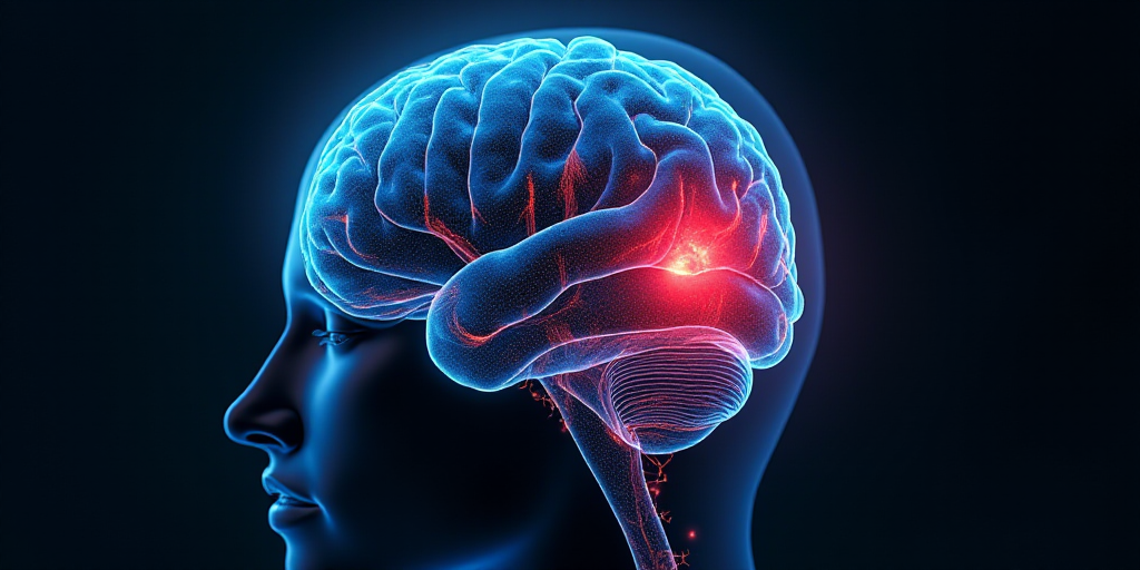
Hidden at the base of the skull, the cerebellum is a structure that accounts for only about 10% of the total volume of the brain, yet it houses more than 50% of all neurons in the central nervous system. This remarkable neuronal density underscores its functional importance, which extends beyond regulating movements and maintaining balance—the traditional roles it has been attributed.
Beyond Movement Regulation
In the past two decades, it has been revealed that the cerebellum is not merely a mechanical regulator but also a modulator of more complex mental processes.
The cognitive-affective cerebellar syndrome (CCAS), first described in 1998, combines motor alterations with difficulties in planning, language, working memory, and emotion regulation. It is believed that the uniformity of the cerebellum’s microarchitecture—which calculates movement adjustments—is also employed to generate “internal models” that anticipate and fine-tune the dynamics of thoughts and emotional states.
There are more examples of the cerebellum’s involvement in other diseases, such as Alzheimer’s. This neurodegenerative disease is characterized by the abnormal accumulation of β-amyloid protein and causes thinning of a layer of neurons called the granular layer, crucial for processing information that reaches the cerebellum. These changes can affect the coordination of mind and body.
In autism, alterations in Purkinjee cells and cerebellar-cortical connections have been observed, contributing to difficulties in interpreting gestures, sounds, or voice tones, impacting communication and socialization.
In schizophrenia, the lack of coordination between the cerebellum and frontal lobe can result in disorganized thoughts or difficulties regulating emotions, as the cerebellum no longer “fine-tunes” these internal processes with the same precision.
From Leonardo to Ramón y Cajal
Long before these discoveries, the cerebellum was a subject of scientific interest and artistic inspiration for two brilliant minds.
Leonardo Da Vinci (1452–1519), the prototype of the Renaissance scientist, was a man who embodied the third culture that did not distinguish between art and science. In his quest to find a relationship between the microcosm and macrocosm, he early on recognized the crucial role of vision and the brain’s integration capacity.
In his anatomical studies, he was interested in the cerebral ventricles and developed a method to inject hot wax into bull skulls, obtaining precise models of the ventricular system. Although it is attributed to the Florentine genius that he named “cerebellum” (from the Latin cerebellum, meaning “small brain,” in reference to its structural similarity to the brain), this claim cannot be verified.
What is known is that his interest was piqued, as he placed the vermis cerebellum (the central part of the cerebellum) in his drawings as a “valve” that regulates the passage between “common sense” and “memory.” This fusion of neoplatonic vision—seeking the soul’s seat—with anatomical rigor allowed him to break from medieval tradition and lay the groundwork for functional anatomy.
Centuries later, Santiago Ramón y Cajal (1852–1934), the father of modern neuroscience, took anatomical art to the cellular level. Through rigorous observation and refinement of staining techniques—particularly the improvement of Camillo Golgi’s silver impregnation method—Cajal clearly visualized the intimate structure of the nervous system.
His gaze focused particularly on the cerebellum, where he discovered and meticulously depicted the complex arborization of Purkinjee cells, one of the most notable for their size and dendritic ramification. In addition to revealing the individuality of neurons, his work laid the foundation for the “neuron doctrine” principle, challenging the dominant idea of a continuous nervous network.
These observations, captured in dozens of drawings that combine scientific rigor with striking artistic value, enabled understanding of the cerebellum not as an isolated organ but as a key structure in motor coordination and sensory processing.
Our Artistic View of the Cerebellum
At the University of Polytechnic Valencia, we have developed a software called DeepCERES from MRI data that allows for the quantification (segmentation) of 27 cerebellar structures at ultra-high resolution using artificial intelligence techniques. This will advance research on this crucial structure.
Moreover, thanks to DeepCERES, we can generate detailed 3D reconstructions of each cerebellar structure, especially the white matter’s inner substance known as “arbor vitae” (tree of life), named for its tree-like shape with multiple branches. Following Leonardo and Cajal’s footsteps, our goal is to capture the complexity and beauty of this hidden gem of the brain.


